
Understanding the respiratory system’s structures and functions is crucial; it facilitates gas exchange, regulates pH, impacts blood pressure, and provides immune defense.
A. Overview of Function
The primary function of the respiratory system is facilitating gas exchange – bringing oxygen into the body and expelling carbon dioxide. This vital process supports cellular respiration, providing energy for all bodily functions. However, its role extends beyond simple gas exchange. The respiratory system actively participates in maintaining blood pH homeostasis, a critical aspect of overall health.
Furthermore, it contributes to blood pressure regulation and offers a crucial line of non-specific immune defense. The system’s structures, from the nasal passages to the alveoli, work in concert to filter, warm, and humidify incoming air, protecting delicate lung tissues. A comprehensive understanding of these multifaceted functions is essential for appreciating the system’s overall importance.
B. Importance of Studying the Respiratory System
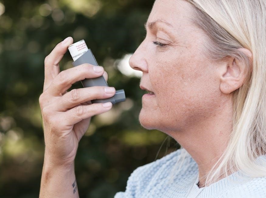
A thorough study of the respiratory system is paramount for healthcare professionals and students alike. Understanding its anatomy and physiology is fundamental to diagnosing and treating a wide range of conditions, from common respiratory infections like pneumonia and bronchitis to chronic diseases such as asthma and COPD. Knowledge of gas exchange mechanisms is crucial for interpreting blood gas analysis.
Moreover, appreciating the system’s role in pH balance and immune defense provides a broader perspective on overall patient health. Studying respiratory physiology also informs interventions like mechanical ventilation and oxygen therapy. Ultimately, a robust understanding empowers effective clinical decision-making and improved patient outcomes.

II. Anatomy of the Upper Respiratory Tract
The upper respiratory tract encompasses the nose, pharynx, and larynx, initiating the pathway for air, filtering, warming, and preparing it for lower airway transit.
A. The Nose and Nasal Cavity
The nose serves as the primary entry point for air into the respiratory system, initiating the crucial process of air conditioning. Within the nasal cavity, nasal conchae – superior, middle, and inferior – create turbulence, maximizing contact between air and the mucous membrane.
This intricate structure facilitates efficient filtering of particulate matter, preventing irritants from reaching the delicate lower airways. Simultaneously, the rich vascular network warms the incoming air to body temperature, and the mucous membrane humidifies it, preventing drying of the respiratory surfaces.
The frontal sinus, located within the frontal bone, contributes to resonance during speech and may play a role in reducing skull weight. Understanding these anatomical features is fundamental to grasping the nose’s vital role in respiratory health.
Structure and Function of Nasal Conchae
Nasal conchae, also known as turbinates – superior, middle, and inferior – are bony structures covered by a highly vascularized mucous membrane within the nasal cavity. Their convoluted shape dramatically increases the surface area available for air contact; This expanded surface is pivotal for efficient conditioning of inspired air.
Functionally, the conchae create turbulent airflow, ensuring thorough mixing of incoming air with the nasal mucosa. This maximizes the processes of filtering airborne particles, warming the air to body temperature, and humidifying it to prevent damage to the lower respiratory tract.
The superior conchae primarily contribute to olfaction, while the middle and inferior conchae are more involved in air conditioning. Their structure is essential for optimal respiratory function.
Role in Filtering, Warming, and Humidifying Air

The upper respiratory tract, particularly the nasal cavity, expertly prepares inhaled air for the delicate lungs. Filtering occurs via nasal hairs and mucus, trapping larger particles and pathogens before they reach deeper airways. Cilia then sweep this debris towards the pharynx for removal.
Warming is achieved through the rich vascular network within the nasal mucosa, raising the air temperature closer to body temperature. Simultaneously, humidification saturates the air with water vapor, preventing drying and damage to the lower respiratory epithelium.
These processes are vital for maintaining optimal lung function and protecting against irritation and infection, ensuring efficient gas exchange.
B. The Pharynx (Throat)
The pharynx, commonly known as the throat, serves as a crucial passageway for both air and food. It’s strategically divided into three regions: the nasopharynx, oropharynx, and laryngopharynx, each with distinct functions. The nasopharynx lies posterior to the nasal cavity, primarily involved in respiration.
The oropharynx continues the pathway for both air and food, while the laryngopharynx leads to the esophagus and larynx. Functionally, the pharynx plays a vital role in swallowing, directing food towards the esophagus, and contributes to speech production by resonating sound.
Its coordinated muscular actions ensure efficient and safe passage of substances.
Divisions of the Pharynx: Nasopharynx, Oropharynx, Laryngopharynx
The pharynx is distinctly segmented into three continuous sections. The nasopharynx, superiorly located, connects to the nasal cavity and contains the adenoids and auditory tubes, functioning primarily in respiration; Inferior to it lies the oropharynx, the central region shared by both respiratory and digestive pathways, including the tonsils.
Finally, the laryngopharynx, the most inferior portion, extends to the larynx and esophagus. This section directs food and air appropriately. Each division exhibits unique anatomical features and contributes specifically to the overall function of the pharynx in breathing, swallowing, and speech.
Function in Swallowing and Speech
The pharynx plays a vital dual role in both swallowing and speech production. During swallowing, its muscular walls contract to propel food towards the esophagus, preventing aspiration into the airway. This complex process requires coordinated muscle action and precise timing.
For speech, the pharynx acts as a resonating chamber, modifying sound produced by the larynx. Adjustments to its shape and size influence the quality and clarity of vocalizations. The coordinated interaction between the pharynx, larynx, and oral cavity is essential for articulate speech.
C. The Larynx (Voice Box)
The larynx, commonly known as the voice box, is a complex structure composed of several cartilages, including the thyroid, cricoid, and epiglottis. The thyroid cartilage forms the prominent Adam’s apple, while the cricoid cartilage sits below it, completing the ring-like structure. The epiglottis acts as a protective flap, preventing food from entering the trachea during swallowing.
Its primary function is sound production, achieved through the vibration of vocal cords within the larynx. Air passing over these cords causes them to vibrate, creating sound. The pitch and volume of the sound are controlled by adjusting the tension and airflow.
Structure of Cartilages (Thyroid, Cricoid, Epiglottis)
The larynx’s structural integrity relies on a framework of cartilages. The largest, the thyroid cartilage, forms the anterior and lateral walls, creating the visible laryngeal prominence – often called the Adam’s apple. Inferior to the thyroid cartilage is the cricoid cartilage, a complete ring that provides attachment for other structures.
The epiglottis, a leaf-shaped cartilage, is crucial for preventing aspiration. It covers the laryngeal inlet during swallowing, directing food and liquids into the esophagus. These cartilages, connected by ligaments and membranes, ensure both airway protection and flexibility for vocalization.
Role in Sound Production
The larynx is paramount in sound production, functioning as the voice box. Air expelled from the lungs passes through the larynx, causing the vocal folds (within the larynx) to vibrate. The tension and length of these folds, controlled by laryngeal muscles, determine pitch.
Louder sounds result from increased airflow and greater vocal fold compression. Resonance chambers – the pharynx, oral cavity, and nasal cavity – amplify and modify the sound, shaping distinct speech patterns. This intricate process transforms exhaled air into articulate vocalizations, essential for communication.
III. Anatomy of the Lower Respiratory Tract
This section details the trachea, bronchi, bronchioles, and lungs – vital components for air conduction and gas exchange, ensuring oxygen reaches the alveoli.
A. The Trachea (Windpipe)
The trachea, commonly known as the windpipe, is a crucial conduit for air traveling between the larynx and the bronchi. Its structure is characterized by C-shaped cartilaginous rings, providing essential support to prevent collapse during inhalation and exhalation.
These rings aren’t complete circles, allowing the esophagus to expand during swallowing. The trachea’s primary function is air conduction, efficiently transporting oxygen-rich air to the lungs and facilitating the removal of carbon dioxide. The inner lining is covered with cilia and mucus-producing cells, forming a mucociliary escalator.
This mechanism traps debris and pathogens, moving them upwards to be expelled, thus protecting the lower respiratory tract. Understanding the trachea’s anatomy and function is fundamental to comprehending the entire respiratory process.
Structure and Cartilaginous Rings
The trachea’s defining structural feature is its series of C-shaped cartilaginous rings. These rings, composed of hyaline cartilage, provide robust support, preventing the airway from collapsing under negative pressure during breathing. The incomplete nature of the ‘C’ shape allows the flexible esophagus to expand when swallowing food.
These rings are connected by fibroelastic ligaments, offering both rigidity and some degree of flexibility. The cartilage maintains airway patency, while the posterior wall, lacking cartilage, allows for esophageal distension. This unique design is vital for efficient air conduction and safe swallowing.
Understanding this structural arrangement is key to grasping how the trachea effectively functions as a stable yet adaptable airway.
Function in Air Conduction
The trachea serves as a crucial conduit for air, efficiently transporting it from the larynx to the bronchi. Its primary function is to ensure unimpeded airflow to facilitate respiration. The smooth inner lining minimizes resistance, allowing for optimal air movement during both inspiration and expiration.
The cartilaginous rings maintain the trachea’s open structure, preventing collapse and ensuring continuous airflow, even with changes in pressure. This consistent pathway is essential for delivering oxygen to the lungs and removing carbon dioxide from the body.
Effective air conduction is paramount for sustaining life, and the trachea’s design perfectly supports this vital physiological process.
B. The Bronchi and Bronchioles
The bronchi, branching from the trachea, initiate the airway’s division into the lungs – primary, secondary, and tertiary structures ensuring widespread distribution. These progressively smaller airways conduct air deeper into the lung tissue, maximizing surface area for gas exchange.
Bronchioles, lacking cartilage, rely on smooth muscle for structural support and airflow regulation. Contraction and relaxation of this muscle control airway diameter, influencing resistance and ventilation. This dynamic control allows the body to adjust airflow based on metabolic demands.
This branching network effectively delivers air to the alveoli, the sites of crucial oxygen and carbon dioxide exchange.

Primary, Secondary, and Tertiary Bronchi
The primary bronchi, originating from the trachea’s bifurcation, serve as the main entry points into each lung – the right and left bronchus. These are substantial airways, maintaining relatively large diameters for efficient airflow.
Secondary bronchi, also known as lobar bronchi, branch off the primary bronchi, each supplying a specific lobe of the lung. The right lung typically has three lobes, corresponding to three secondary bronchi, while the left lung has two lobes and two secondary bronchi.
Tertiary bronchi, or segmental bronchi, further divide from the secondary bronchi, supplying individual segments within each lobe. This hierarchical branching ensures comprehensive air distribution throughout the lungs.
Bronchiole Structure and Smooth Muscle Control
Bronchioles represent smaller airways branching from the tertiary bronchi, lacking cartilage support but possessing a significant amount of smooth muscle within their walls. This smooth muscle plays a critical role in regulating airway diameter, influencing airflow resistance.
Smooth muscle contraction causes bronchiole constriction, decreasing airflow, while relaxation leads to bronchodilation, increasing airflow. This control is largely mediated by the autonomic nervous system and local chemical signals.
The degree of smooth muscle control allows the body to adjust ventilation to match metabolic demands, ensuring adequate oxygen delivery and carbon dioxide removal. Alterations in this control contribute to respiratory conditions like asthma.
C; The Lungs
The lungs, the primary organs of respiration, are cone-shaped structures located within the thoracic cavity. The right lung is divided into three lobes – superior, middle, and inferior – while the left lung has two lobes – superior and inferior – accommodating the heart.
These lobes are further subdivided into smaller units called bronchopulmonary segments, each supplied by its own bronchus and blood vessels. Within the lungs reside millions of tiny air sacs called alveoli, the sites of gas exchange.
Alveoli are surrounded by a dense network of capillaries, facilitating the transfer of oxygen into the bloodstream and carbon dioxide out.
Lobes of the Lungs (Right vs. Left)
The lungs aren’t symmetrical; the right lung possesses three lobes – superior, middle, and inferior – clearly demarcated by fissures. This arrangement maximizes surface area for gas exchange. Conversely, the left lung has only two lobes: superior and inferior.
This difference in lobation is due to the presence of the heart, which extends towards the left side of the chest, creating space constraints. The cardiac notch on the left lung accommodates this anatomical feature.
Understanding this structural variation is vital for interpreting chest radiographs and diagnosing pulmonary conditions, as lobe-specific pathologies can be identified.
Alveoli: Structure and Gas Exchange
Alveoli are tiny, balloon-like air sacs within the lungs, representing the primary site of gas exchange. Their incredibly thin walls, composed of a single layer of epithelial cells, facilitate efficient diffusion of oxygen and carbon dioxide.
Surrounding each alveolus is a dense network of capillaries, ensuring close proximity between air and blood. This structural arrangement maximizes the surface area available for gas exchange – estimated to be around 70 square meters in humans!
Oxygen diffuses from the alveoli into the blood, while carbon dioxide moves from the blood into the alveoli to be exhaled, driven by concentration gradients.

IV. Physiology of Respiration
Respiration involves breathing mechanics – inspiration and expiration utilizing muscles – alongside crucial gas exchange between lungs, blood, and cells for life support.
A. Mechanisms of Breathing
Breathing, or ventilation, is driven by pressure differences created by the diaphragm and intercostal muscles. Inspiration occurs when these muscles contract, expanding the thoracic cavity and decreasing intrapulmonary pressure, drawing air into the lungs. Conversely, expiration typically involves relaxation of these muscles, reducing thoracic volume and increasing pressure, forcing air out.
Lung volumes – tidal volume, inspiratory reserve volume, expiratory reserve volume, and residual volume – contribute to overall lung capacity. Capacities, like vital capacity and total lung capacity, are combinations of these volumes, providing insights into respiratory function and potential limitations. Understanding these mechanics is fundamental to grasping respiratory physiology and identifying potential abnormalities.
Inspiration and Expiration: Muscles Involved
Inspiration primarily relies on the diaphragm, contracting to increase thoracic cavity volume, and the external intercostal muscles, elevating the rib cage. These actions lower intrapulmonary pressure, facilitating air intake. Expiration is generally passive, utilizing the elastic recoil of the lungs and relaxation of inspiratory muscles.
However, forced expiration engages internal intercostal muscles, depressing the rib cage, and abdominal muscles, increasing intra-abdominal pressure. Accessory muscles, like sternocleidomastoid and scalenes, assist during labored breathing. Comprehending the roles of these muscles is vital for understanding normal respiratory function and identifying muscular imbalances impacting breathing efficiency.
Lung Volumes and Capacities
Lung volumes represent distinct compartments of air within the lungs: tidal volume (TV), inspiratory reserve volume (IRV), expiratory reserve volume (ERV), and residual volume (RV). Lung capacities combine two or more volumes, including vital capacity (VC – TV + IRV + ERV), functional residual capacity (FRC – ERV + RV), and total lung capacity (TLC – VC + RV).
Understanding these measurements is crucial for assessing respiratory health. Reduced volumes or capacities can indicate restrictive lung diseases, while altered ratios suggest obstructive conditions. Spirometry, a common pulmonary function test, measures these parameters, aiding in diagnosis and monitoring disease progression. Accurate interpretation requires knowledge of normal values and potential influencing factors.
B. Gas Exchange
Gas exchange, vital for life, occurs in two phases: external and internal respiration. External respiration involves oxygen moving from the alveoli into the blood, and carbon dioxide moving from the blood into the alveoli, driven by partial pressure gradients. This happens across the respiratory membrane.
Internal respiration is the exchange of gases between the blood and body tissues. Oxygen diffuses from the blood into cells, while carbon dioxide moves from cells into the blood. Efficient gas exchange relies on adequate ventilation, perfusion, and a thin respiratory membrane. Understanding these processes is key to grasping respiratory physiology.
External Respiration (Lungs to Blood)
External respiration, the initial stage of gas exchange, happens within the lungs; Oxygen diffuses across the alveolar and capillary walls, moving from the alveoli – where oxygen concentration is high – into the pulmonary capillaries, where it’s lower. Simultaneously, carbon dioxide moves in the opposite direction.
This diffusion is governed by partial pressure gradients; gases flow from areas of high pressure to low pressure. The efficiency of this process depends on the respiratory membrane’s thinness and large surface area. Adequate ventilation and perfusion are also crucial for optimal oxygen uptake and carbon dioxide removal.
Internal Respiration (Blood to Cells)
Internal respiration represents the gas exchange between the blood and body tissues. Oxygen diffuses from the capillaries, where its concentration is higher, into the interstitial fluid and then into cells, where it’s utilized for cellular metabolism. Conversely, carbon dioxide, a waste product of metabolism, moves from the cells into the blood.
Like external respiration, this process relies on partial pressure gradients. The efficiency is affected by factors like blood flow, tissue metabolism, and the distance between capillaries and cells. This vital exchange delivers oxygen for energy production and removes carbon dioxide, maintaining cellular function and overall homeostasis.

V. Control of Respiration
Breathing regulation involves the brainstem, specifically the medulla oblongata and pons, alongside chemoreceptors that monitor blood gas levels for adjustments.
A. Role of the Brainstem (Medulla Oblongata and Pons)
The brainstem, comprising the medulla oblongata and pons, serves as the primary respiratory control center. The medulla contains the dorsal respiratory group (DRG) and ventral respiratory group (VRG), crucial for establishing the basic rhythm of breathing. The DRG primarily controls inspiration, while the VRG manages both inspiration and expiration, especially during forceful breathing.
Furthermore, the pons houses the pneumotaxic and apneustic centers, modulating the medulla’s activity. The pneumotaxic center limits inspiration, shortening the breathing cycle, and the apneustic center prolongs inspiration. This intricate interplay ensures appropriate ventilation rates based on the body’s metabolic demands, demonstrating the brainstem’s vital role in maintaining respiratory homeostasis.
B. Chemoreceptors and Regulation of Breathing Rate
Breathing rate is finely tuned by chemoreceptors that monitor blood gas levels. Central chemoreceptors, located in the medulla oblongata, primarily detect changes in pH caused by carbon dioxide (CO2) levels. Increased CO2 leads to a decrease in pH, stimulating increased ventilation to expel excess CO2.
Peripheral chemoreceptors, found in the carotid and aortic bodies, respond to changes in oxygen (O2), CO2, and pH. While less sensitive to CO2 than central receptors, they are crucial for detecting significant drops in O2 levels. These receptors relay information to the brainstem, adjusting breathing rate and depth to maintain optimal blood gas homeostasis.